
Baktybek Kojonazarov is appointed Head of the Small Animals Imaging Platform at the Institute for Lung Health (ILH), Justus Liebig University Giessen (JLU), Germany.
Baktybek Kojonazarov was born in Frunze, Kyrgyz Republic. He has successfully graduated from Kyrgyz State Medical Academy and specialised in the field of internal medicine and cardiology. Several years he worked with patients suffered from pulmonary hypertension and heart diseases in the National Center of Cardiology and Internal Medicine and at the same time studied genetic and molecular mechanisms of high altitude pulmonary hypertension in the Institute of Molecular Biology and Medicine, Bishkek, Kyrgyzstan. He then joined in 2001-2002 laboratory of Professor Robert Naeije and department of cardiology of Professor Philippe van de Borne at Erasme Hospital, Free University of Brussels with the support of the Baron Seutin’s grant and investigated the role of autonomous nervous system in patients with heart failure. He has successfully defended his PhD in the field of medical science in 2005. In 2009 as an expert in echocardiography, he was invited to join laboratory of Pulmonary pharmacotherapy at the JLU, Giessen, Germany. As an expert in the field of imaging, cardiovascular, respiratory physiology and pathophysiology, he has established several imaging techniques and approaches in the University of Giessen for in vivo and ex vivo evaluation of lung and heart perfusion, cardiac and lung function, lung density, aeration and in vivo fluorescence molecular imaging in rodents. In 2020, he was appointed Head of Small Animal Imaging platform at the ILH, Giessen.
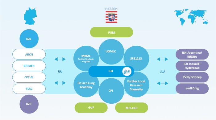
He also serves as the Principal Investigator at the German Center for Lung Research (DZL), faculty member of the Universities of Giessen and Marburg Lung Center (UGMLC), adjunct faculty of the Excellence Cluster Cardio-Pulmonary Institute (CPI). Moreover, he is leading central project (CP02) within Collaborative Research Center (CRC1213) – “Pulmonary hypertension and cor pulmonale”. Dr. Kojonazarov published 58 research articles (Date: 15th Jan 2021) in well-renowned scientific journals such as Nature Medicine, Science Translational Medicine, Nature Communications, Nature Metabolism, Circulation, Circulation Cardiovascular Imaging, American Journal of Respiratory and Critical Care Medicine, European Respiratory Journal, American Journal of Respiratory Cell and Molecular Biology, Cardiovascular Research.
Dr. Kojonazarov aims to develop and establish novel in vivo imaging biomarkers in animal models of the pulmonary (pulmonary fibrosis, COPD, lung cancer) and cardiovascular diseases (pulmonary hypertension, heart failure).
In recent years, imaging modalities have become indispensable tools in the basic and preclinical research. The rapidly developing high-resolution in vivo imaging technologies provide an opportunity for studying biological processes of living organisms. State of the art non-invasive small-animal imaging modalities provide qualitative and quantitative anatomical and functional information, with possibility to perform longitudinal studies allowing monitoring the disease progression. We are convinced that non-invasive imaging techniques allows us to reduce the number of animals in our research investigation according to the 3R principle (reduction, refinement, replacement). In our department we are using the most suitable imaging modalities for in vivo small-animal imaging such as ultrasonography, microscopic computed tomography (µCT) and 3D fluorescence molecular tomography (FMT) with unique approaches and methods to detect pathological processes up to microscopic level.
Overview of methods/techniques provided by Small Animal Imaging Platform
Small Animal Imaging Platform provides highly comprehensive and standardized expertise in screening, phenotyping and detailed characterization of the animal models of respiratory and cardiovascular diseases with established noninvasive imaging techniques.
 Echocardiography (Ultrasound bio-microscopy) – imaging platform is operated with Vevo3100 high-resolution ultrasound imaging system (Visaulsonics, Canada). To perform acquisition and analyses of the cardiac morphology and function in rodents from embryos to neonates and adult. Myocardial and vascular strain and strain rate analyses of the left and right ventricles, large and small arter
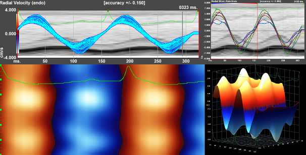 ies.
ies.
Comprehensive analyses of the segmental cardiac function, intra- and interventricular dyssynchrony in rodents with various cardiovascular pathologies. Analyses of the vascular pathologies and quantitative analyses of the vessels wall motions, vessels stiffness by PWD, vessels microanatomy and intima-media thickness (IMT).
Microscopic Computed Tomography (µCT) – Quantum GX high resolution imaging system
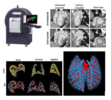
High resolution (up to 5 µm), high speed (8 sec), low dose x-ray imaging is suitable for longitudinal in vivo studies. Detailed analyses of the airways and lung (volume, diameter, density, aeration, functional residual capacity (FRC), tissue compartment) morphology. Analyses of the heart morphology and function (end-diastolic, end-systolic volumes, stroke volume, cardiac output, ejection fraction), including multiphase reconstruction and detailed functional analyses of the systolic and diastolic functional parameters. In vivo (lungs blood volume) and ex vivo (volume of arterial and venous systems, branches, junctions)lung and heart (coronary arteries tree) perfusion with detailed analyses of the vascular tree. µCT-derived virtual bronchoscopy useful tool for 3D visualization of the bronchial tree and diagnostic of lung pathologies including lung cancer, bronchoectases etc. .
3D Fluorescence Molecular Tomography (FMT)
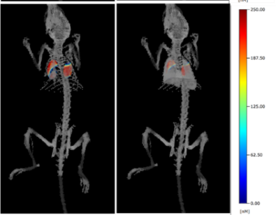
 3D Fluorescence Molecular Tomography (FMT, PerkinElmer, USA) combined with microCT system (Quantum GX, Rigacu, Japan, PerkinElmer, USA) to investigate molecular events in whole body in response to various pathologies. Broad spectrum of near-infrared agents (spectrum 680nm, 750 nm, 800nm) available to study inflammation (MMPs and Cathepsin activities), apoptosis (annexinV), metabolism (2DG), integrin (αvβ3, αvβ5) and carbonic anhydrase IX and XII expressions, as well as vascularity, perfusion and permeability in lung, heart disease and cancer research.
From past several years, Dr Kojonazarov is interested in adaptive and maladaptive pulmonary vascular and heart remodeling, as well as cardio-pulmonary interaction in pulmonary hypertension and various lung diseases. To investigate the above, Dr Kojonazarov established several imaging methods and approaches for detailed characterization of the pulmonary and cardiac function in rodents. Therefore, the small animal imaging platform not only provides service for other departments within ILH, but also focuses on the following major objectives:
Dr. Baktybek Kojonazarov
Small Animal Imaging Platform
Institute for lung health (ILH)
Justus Liebig University Giessen
Aulweg 130
35392 Gießen
Tel: +49 (0) 641 99 46802
Fax: +49 (0) 641 99 46829
Email:
baktybek.kojonazarov@innere.med.uni-giessen.de

![]()